CANINE HIP DYSPLASIA (CHD)
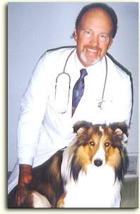
by T. J. Dunn, Jr. DVM
HIP DYSPLASIA in dogs! With this report we'll clear up some misconceptions, describe what CHD is, what effect the condition can have on the dog and what
can be done about it. Because
Hip Dysplasia in dogs is a complex topic, it requires extensive consideration in order to have a good understanding of its nature. Cats do suffer from Hip Dysplasia, too, but it is seen less frequently in cats.
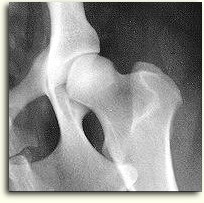
WHAT IS IT: "Hip dysplasia" simply stated means an "abnormal formation" of the hip joint. Think of the condition first as a looseness in a joint that should be snug - then most of the problems attendant to hip dysplasia are a result of this "looseness". See the image on the right a few paragraphs down for an example of a nice, normal, snug hip joint.
The normal anatomy of the hip joint is a classic Ball and Socket joint. The head of the femur (the "Ball") is supposed to match the acetabulum (the "Socket"). A good hip joint has a neat, snug fit between the ball and socket - that is, the head of the femur should not be slipping and slopping around somewhere in the neighborhood of the acetabulum!
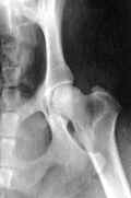
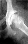
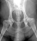
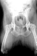
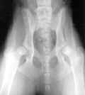
There are infinite variations of dysplasia - ranging from only very slight changes from normal to complete dislocation. (There are a number of examples of actual radiographs in the table near the bottom of this page. Click on any x-ray image to enlarge it.) Consequently, no two dogs will be affected by CHD exactly alike.
HOW IS CHD ACQUIRED? This is one disorder that has been proven, positively, to have a genetic basis. How much of a genetic origin in each case can vary from 25% to 85%. A condition that is completely determined by genetics, for example gender, has a Heritibility Factor of 1. A condition totally unaffected by genetics, for example a broken leg, has a Heritibility Factor of zero.
Studies have shown that CHD's Heritibility factor ranges from .25 to .85; this is a significant genetic contribution. So the Heritibility Factor for a given dog is the result of a combination of the Heritibility Factors from each parent. Simply put . . . if the parents are carrying genetic material for
hip dysplasia - so will the offspring. And the greater the genetic contribution for loose hips or malformed bone or abnormal muscle mass (Heritibility Factor) from
the parents, the greater the chances for hip dysplasia in the offspring.
The expression of hip dysplasia in any dog has other determinants, though; genetics play only a varying role in the total picture. The effect of the developing dog's environment does play a role in the clinical (observable) signs of dysplasia, although just like the genetic component the effects of environment are variable and not completely understood. To illustrate the complexity of the environmental issue, listen to this: It is possible for a dog with known genetic components for hip dysplasia (called genotype) to not show any clinical signs of trouble if the environmental factors are favorable. So the dog can be dysplastic and not show observable signs of it until middle or old age. I have seen this fairly commonly in practice and it is always an important issue with breeders who assume that their dog is normal just because it hasn't shown any signs of hip trouble. Why take pelvic x-rays for dysplasia when the dog has always acted perfectly fit, they reason. There is no excuse for NOT taking pre-breeding x-rays.
I have seen a number of breeders who sold litters of pups where the parents have not been x-rayed for CHD and who were shocked a year or so later when the phone started ringing about "that pup you sold has hip dysplasia". Trust me, it happens. Also, if two dogs that have the same genotype (genetic makeup) are exposed to different environmental conditions, their expression of hip trouble can be quite dissimilar. Little wonder that the topic has such a wide range of information and misinformation regarding it.
Some of the environmental aspects that can affect the observable expression of hip dysplasia are the following:
1.
Nutrition - There are reports that in puppies a restricted calorie intake could restricted the growth rate, and in turn will lessen the potential for the dog to develop hip dysplasia. (I wouldn't suggest doing this to any pup... it makes as much sense as stealing money from your own checking account!) The problem is that some restricted diets restrict the fat and protein content and increase the carbohydrate content of the food. Bad! See a better way in the discussion in ThePetCenter.com
here. The real goal should be to keep growing pups from becoming OVERWEIGHT. Restricting fat and protein in a growing pup can be a disaster. A high quality, meat-based diet is absolutely necessary for growing pups, just don't feed so much of it that the pup becomes overweight.
2.
Physical Activity - In a young, growing dog with a genotype (genetic makeup) for CHD who will eventually develop some trouble because of it, will develop more arthritis and have more eventual difficulty if it is highly active physically. Climbing stairs, jumping into and out of pick-up trucks, running with other normal dogs can all subject the growing hip structures to unwarranted stress and trauma and increase future discomfort for the dog. The effects of this excessive activity is worsened in an overweight pup. (In a normal, growing dog, all these activities will not cause hip dysplasia!)
3.
Bedding - There is no scientific proof,
but lots of observational conclusions, that pups reared especially during the nursing period on slippery surfaces such as newspapers will be prone to hip difficulties. That is not to say that smooth concrete, wood or newspaper surfaces cause dysplasia, just that they can make a bad situation worse. Better surfaces for newborn pups would be blankets or towels... something they can get a better grip on.
MUSCLE AND CHD: Research has shown that dogs with CHD have significantly decreased sizes of total pelvic musculature surrounding and acting on the hip joint. Whether this is a contributing factor or a result of hip dysplasia remains to be proven.
One muscle that can contribute to worsening of hip dysplasia is the Pectineus Muscle. In dogs with a strong genetic background for CHD, the microscopic makeup and contractibility of the Pectineus Muscle are strikingly different from the same muscle of normal dogs. The theory is that a tight or inelastic Pectineus Muscle causes tension in such a direction that the force tends to pull the head of the femur away from the acetabulum. So the tight muscle creates more looseness in the joint. I have had good results in about 50% of the cases I have surgically excised a portion of the Pectineus Muscle. The patients were more comfortable and mobile almost immediately. This Pectineal Myotomy surgery has no effect on the arthritic changes in the hip joints; it can make the dog more comfortable.
LIGAMENT OF THE HEAD OF THE FEMUR: Attaching to the head of the femur from the center of the hip socket is a tough fibrous ligament called the Ligament of the Head of the Femur. If this ligament is stretched or torn, the hip joint will be less stable . . . and this is exactly what happens to dogs with dysplasia. In fact, some of the first changes to take place in young dogs developing hip dysplasia occur in this ligament especially if the muscle mass of the pelvis is underdeveloped. The ligament swells, develops tiny tears and stretches. In advanced CHD this ligament can totally break down and cause more harm than good.
JOINT CAPSULE: This tissue, which if you could hold it, would feel like the wall of a thick balloon It surrounds the joint and produces synovial fluid to nourish and lubricate the joint cartilage. In addition, the joint capsule provides some support to the joint.
In dysplastic joints the capsule becomes irritated, stretched, and scarred. In advanced cases the capsule will lose its elasticity and inhibit a full range of motion in the joint. A large percentage of the pain associated with hip dysplasia originates from inflamed nerve endings in the joint capsule so any pathology here will have a noticeable affect on the dog.
CARTILAGE: The surfaces of the head of the femur and the acetabulum are covered with what is termed hyaline cartilage. In a dysplastic joint the points of pressure and the amount of pressure applied to areas of cartilage surfaces are abnormal. The cartilage is being asked to do things it physically cannot accomplish, so it changes or disintegrates as a response. The changes range from thickening in abnormal areas to thinning in others. Sometimes the pounding it gets erodes the cartilage down to the underlying bone! The outcome is more pain and discomfort, more inflammation, more calcium deposits from inadequate healing attempts and eventual breakdown of the joint as a unit. Nutriceuticals such as Chondroitin Sulfate and Glucosamine may be effective in aiding the repair and maintenance of this articular cartilage.
BONE CHANGES: Since bone is alive it responds to stress and grows in a manner that tends to distribute weight loads evenly. As a result of posture changes brought on by discomfort, the dog's weight bearing forces stress the bone in
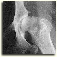
unnatural ways. The bone does what it is supposed to do as a response and changes its shape. The bone doesn't know doesn't know that the shape it changes to is abnormal.
Ultimately, this abnormal shape to the thigh bone and acetabulum create more difficulty with stability and a vicious cycle ensues that spells trouble for the dog. See the images below for a comparison of before/after bony changes. The final outcome of bony remodeling in unstable hip joints is
Degenerative Joint Disease.
SIGNS OF CHD IN YOUNG DOGS: What you will see first is a pup that runs with both back legs nearly together, almost like a rabbit would run. After exercise the pup will be reluctant to rise, will sit back as if unsteady and will have difficulty climbing stairs or inclines. The pup might look slightly underdeveloped in the rear quarters. When it stands the rear legs may not be parallel, but rather too near each other at the hocks (ankles) called "cow hocked".
You might notice a boniness to the pelvic area from lack of good muscle development. Another hint of trouble is an inability to extend the leg backward very far (decreased range of motion). Note: Many pups rest or sleep in a frog-like position with knees extended out to either side - this is a good sign and shouldn't alarm you.
In severe cases of dysplasia, the young dog will rock forward to support more weight on the front legs (which can create trouble in the shoulders and elbows). When dogs do this it seems as if they are tip-toeing or walking very lightly on their rear legs. A dysplastic pup will be reluctant to jump or "stand up" on its hind legs. Signs usually being between five and eights months of age. But remember, as we learned above, some dogs do not show any signs at all of hip joint degeneration until mature adults.
AN INTERESTING CASE: Here is a classic example of why it is so important to take a radiograph of the sire and bitch prior to 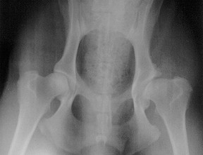 breeding. In this example the owner had a two year old male Golden Retriever that was totally healthy by any observable standards. It ran, jumped, swam and had never showed any kind of lameness. The owner had the dog x-rayed and guess what? The film displayed severe abnormalities in the left hip joint. Were the changes due to a genetic propensity for hip joint abnormalities? Or was this actually due to an injury early in the pup's life that impacted the proper growth of the joint structures? No one can say for certain. But IF the abnormal hip was due to genetic determiners why take a chance that, if bred, the litter might have even worse hip joint conformation? The owner decided not to breed the dog.
SIGNS OF CHD IN OLDER DOGS: Some dogs with dysplasia escape pain or simply accept it as a fact of life and don't complain until degenerative joint disease sets in. Affected dogs will sit rather than stand, have trouble arising, run with the rear legs together and not be able to keep up any more on Sunday walks. Every veterinarian has been mystified on occasions where an x-ray of an older dog, who only recently seemed to be having hip trouble, reveals extensive degenerative changes in the hips due to long term dysplasia.
It is very important to keep this fact in mind: A dog can appear normal and yet have hip dysplasia. Just because a four-year-old dog isn't showing signs of trouble is not sufficient evidence to state "it couldn't possibly have hip dysplasia". I have heard supposedly responsible breeders make that statement and it takes lots of convincing to get them to believe otherwise.
If you are involved with a breed in which CHD has been reported, and you wish to improve the breed as well as have happy owners of your pups you must know if your breeding stock is prone to CHD. And neither you, your cousin, the mailman OR your veterinarian can tell if your dog has CHD unless some basic guidelines are followed.
DETERMINING THE PRESENCE OF CHD: Dogs with obvious signs of CHD (hip soreness, difficulty arising or climbing inclines, muscle atrophy over the rump, limping) are not a challenge to confirm as such. So this discussion will apply more to the dog that seems to be normal but you are either not sure or need to know for breeding or training/working reasons. The minimum data required is a pelvic x-ray taken under anesthesia . . . PERIOD! You MUST have the x-ray to know if the dog is normal!
PennHIP: (University of
Pennsylvania Hip Improvement Program) See an entire article about PennHIP
here.
Commercially available since 1993, this procedure has been and was developed as an
objective method of evaluating dogs’ hip structure. It evolved as a direct result of the subjectivity factors and age constraint (maturity) limitations inherent to evaluation and certification of dogs by the OFA and other screening programs. PennHIP research published in peer reviewed journals has shown that different breeds have different susceptibility to osteoarthritis. Therefore, in PennHIP evaluations each breed is compared to its own. Only PennHIP certified veterinarians can do the PennHIP evaluation but many veterinarians are becoming certified in this procedure.
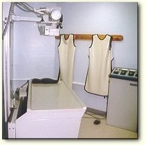
Why is anesthesia required in order to have the dog radiographed? To have an x-ray that yields the information you're trying to discover the dog must be perfectly relaxed. Because the position required to take a diagnostic x-ray is a somewhat unnatural one, even very gentle,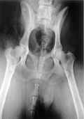 cooperative dogs cannot relax enough to be x-rayed properly. See the x-ray on the right. It is a great example of poor positioning. (Click on it to enlarge.) The dog is tilted to its left (to our right) and the view we see of the structures is imbalanced. One hip looks OK and the other bad but in reality this view is worthless. Nothing is more frustrating for the veterinarian than to have an owner say "I need to know if this dog has any signs of hip dysplasia. Take an x-ray, but I don't want you to use anesthesia; this dog will do anything you tell it to do, so an anesthetic isn't necessary." Unless at the time of exposure of the x-ray, the dog is positioned precisely, with no movement, the x-ray will not be credible. You won't get the information you need! Your veterinarian will look for an x-ray image that looks like the nice, normal hip at the beginning of this article.
Another great advantage of anesthesia is that the veterinarian can only then palpate and manipulate the hips to actually feel the degree of looseness. Also, the tension of the Pectineus Muscle is best assessed under anesthesia. Any grating or grinding from calcium deposits along the hip joints can be evaluated better than attempting to do so on an awake patient. If you need the information, the dog needs the anesthetic.
If the pelvis is tipped only slightly to one side or the other, one hip can appear normal that isn't and one can appear dysplastic that isn't! To complicate things, 10% of dysplastic dogs will be affected in only one hip! Better do the x-ray right!
The importance of radiography cannot be overstated. It can be done early, say five or six months of age, if dysplasia is suspected. If the results are questionable, reserve breeding until a time when the x-rays are conclusive. Generally, by the time the dog is full grown the x-rays will properly reveal the status of the hips. The OFA (OFA.org) will not classify hips in dogs until they are two years of age. The OVC will certify hips at 18 months of age.
The advantage of radiography in a younger animal is that if you plan on breeding it you can eliminate fruitless time and financial and emotional expense related to breeding if the x-rays show unquestionable hip dysplasia. There have been many disappointed, depressed dog owners whose expectations for breeding were high and were shocked back to reality when their two-year-old dog showed x-ray evidence of dysplasia... two years of planning, training and dreams of great litters down the tube. If only the parents had been x-rayed. If only preliminary x-ray were taken eighteen months ago. Again, the advantage of the PennHIP procedure is obvious since dog over 4 months of age cane be evaluated.
It is very sad indeed for any pet owner to see their special pal affected by the discomfort and mobility problems associated with Canine Hip Dysplasia. Fortunately, armed with knowledge and forethought, highly selective breeding is your best defense against CHD.
I have seen a number of breeders who sold litters of pups where the sire and bitch had not been x-rayed for CHD. The breeders were shocked a year or so later when the phone started ringing and upset dog owners complained because "that pup you sold us has hip dysplasia". |
For your inspection you can click on any of the images below to see
a full sized photo in a new window.
 |
 |
 |
 |
 |
| Normal Hip Joint |
Severe Hip Dysplasia and Degenerative Joint Disease |
Dysplastic in both hips, one is subluxated |
Osteoarthritic hips due to hip dysplasia |
These hips are almost dislocated they are so loose fitting |
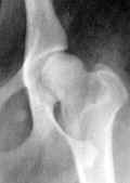 |
 |
 |
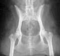 |
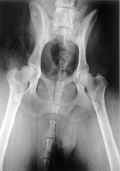 |
| Moderately dysplastic hip |
Crippling arthritis |
Not bad but not good either |
A normal cat hip image |
Poor positioning for an accurate diagnosis |
Please note that if you show an x-ray of a dog's hips to a veterinarian, the evaluation will be subjective. If there is a disagreement regarding a diagnosis, it is best to get more than one opinion.

 can be done about it. Because Hip Dysplasia in dogs is a complex topic, it requires extensive consideration in order to have a good understanding of its nature. Cats do suffer from Hip Dysplasia, too, but it is seen less frequently in cats.
 the parents, the greater the chances for hip dysplasia in the offspring.
 but lots of observational conclusions, that pups reared especially during the nursing period on slippery surfaces such as newspapers will be prone to hip difficulties. That is not to say that smooth concrete, wood or newspaper surfaces cause dysplasia, just that they can make a bad situation worse. Better surfaces for newborn pups would be blankets or towels... something they can get a better grip on.
 unnatural ways. The bone does what it is supposed to do as a response and changes its shape. The bone doesn't know doesn't know that the shape it changes to is abnormal.


